Tipping points in the microcosmos

How the microverse regulates life and the environment

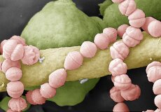
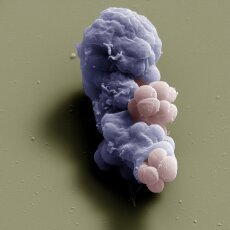
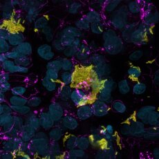
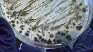
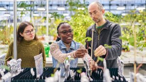
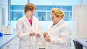
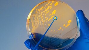
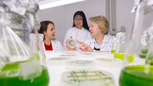
They are the tiniest organisms on the planet—but also the most numerous and the most successful. Microorganisms were the first living things on Earth. But that’s not all: over millions of years, they have shaped our planet, its biosphere, the atmosphere, and all higher forms of life. Consequently, the environment, the climate, and the health of plants, animals, and humans depend on microorganisms and their complex communities. Based in Jena, the Cluster of Excellence »Balance of the Microverse« examines how the microverse—the sum of all microbial communities—regulates environmental and vital processes.

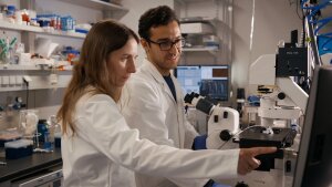
Researchers from the fields of microbiology, chemical ecology, biogeochemistry, photon technology and infection biology are investigating how microbial communities function, how resilient they are, and what happens when microbiomes reach »tipping points«.

Editorial »Tipping points in the microcosmos«

Pagination Page 1 of 126

Gallery

  • This coloured scanning electron microscopy image shows a co-culture assay of the yeast fungi Candida albicans (hypha in yellow, conidium in green) and the bacterium Enterococcus faecalis (rose). These microbes can form a biofilm, including on cardiac catheter tubes, and cause health problems for patients. The hyphae have a diameter of approx. 1.5 micrometres, while the conidia measure roughly four micrometres. The bacterial cells are around half a micrometre in size.
    This coloured scanning electron microscopy image shows a co-culture assay of the yeast fungi Candida albicans (hypha in yellow, conidium in green) and the bacterium Enterococcus faecalis (rose). These microbes can form a biofilm, including on cardiac catheter tubes, and cause health problems for patients. The hyphae have a diameter of approx. 1.5 micrometres, while the conidia measure roughly four micrometres. The bacterial cells are around half a micrometre in size.
    Image: Sandor Nietzsche (Elektronenmikroskopisches Zentrum des UKJ)
  • This coloured scanning electron microscopy image shows a phagocytic cell, part of the immune system in the pulmonary alveoli (alveolar macrophage, purple), which engulfs Staphylococcus aureus bacteria (rose) and renders them harmless. This image was produced in a lab-based experiment to investigate age-related deterioration in immune defence. Staphylococcus aureus can trigger severe pulmonary diseases. These bacteria are roughly one micrometre in diameter.
    This coloured scanning electron microscopy image shows a phagocytic cell, part of the immune system in the pulmonary alveoli (alveolar macrophage, purple), which engulfs Staphylococcus aureus bacteria (rose) and renders them harmless. This image was produced in a lab-based experiment to investigate age-related deterioration in immune defence. Staphylococcus aureus can trigger severe pulmonary diseases. These bacteria are roughly one micrometre in diameter.
    Image: Sandor Nietzsche (Elektronenmikroskopisches Zentrum des UKJ)
  • Polychromatic bacteria. The colours of the marine bacterium Marinobacter alginolytica can be attributed to ordered bacterial cells that form a photonic crystal and produce interference effects (DOI: 10.1073/pnas.2309757121).
    Polychromatic bacteria. The colours of the marine bacterium Marinobacter alginolytica can be attributed to ordered bacterial cells that form a photonic crystal and produce interference effects (DOI: 10.1073/pnas.2309757121).
    Image: Colin Ingham
  • This image, taken using fluorescence microscopy, shows a crosssection of the large intestine of a mouse infected with Candida albicans. The thread-like and yeast-like fungi (yellow) appear in the mouse’s faeces. The intestinal epithelial cells are shown in blue, with the mucus produced by the cells shown in magenta.
    This image, taken using fluorescence microscopy, shows a crosssection of the large intestine of a mouse infected with Candida albicans. The thread-like and yeast-like fungi (yellow) appear in the mouse’s faeces. The intestinal epithelial cells are shown in blue, with the mucus produced by the cells shown in magenta.
    Image: Merle Hammer
  • Vibrio cholerae bacteria (yellow) have infected human intestinal cells (cyan) growing on a biochip. The pathogenic bacteria are capable of disrupting the junctions between the cells (magenta).
    Vibrio cholerae bacteria (yellow) have infected human intestinal cells (cyan) growing on a biochip. The pathogenic bacteria are capable of disrupting the junctions between the cells (magenta).
    Image: Adrian Feile