Ultra-sensitive surgical robots
An interdisciplinary research team from the University and the University Hospital is developing a sensor-based support system for tumour operations.
By Ute Schönfelder
An interdisciplinary research team at the University of Jena and Jena University Hospital is working with other project partners to develop a sensor-based support system for tumour surgery. Launched in 2023, the »Sensorized Surgery« project is supported by the Carl Zeiss Foundation through its »Breakthroughs« funding programme. It aims to develop a system that assists surgeons with tumour removal by providing real-time visual and haptic feedback during the operation. In addition to teams from the fields of computer science and medicine, the project also includes researchers from the Leibniz Institute of Photonic Technology and TU Ilmenau.
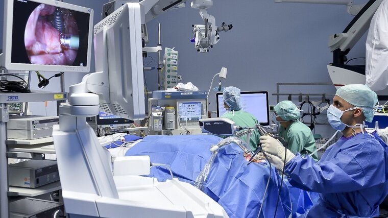
Malignant tumours are often life-threatening and are therefore surgically removed where possible. »This involves removing all of the tumour tissue while conserving the healthy tissue surrounding it,« says Prof. Dr Orlando Guntinas-Lichius of the ENT department at Jena University Hospital, who is also coordinator of the project team. Surgeons need to identify the margin between the tumour and the healthy tissue and make precise incisions accordingly. To date, surgeons have used techniques such as white light video endoscopy before ultimately deciding where to make incisions based on their experience.
»In up to 30% of cases, however, this approach fails to remove the tumour completely, which significantly reduces the patient’s chance of survival,« as Guntinas-Lichius states. Despite the fact that surgical robots can now work with increasing levels of precision, it is not possible to reduce this percentage without higher resolution and clearer views of the tissue margins.
This is exactly what the interdisciplinary team has set out to achieve by developing complex, sensorized surgical methods. Using novel biophotonic imaging technology and biomechanical sensors, the researchers hope to provide high-resolution, real-time outlines of the tumour margins during the operation, with haptic feedback so that surgeons can not only see the tumour but »feel« it, too.
AI plays a central role in this endeavour. It is based on data from histopathological examinations of head-neck and brain tumours, both from Jena University Hospital and later from a trans-regional network of hospitals. This data has been used to train several decentralized machine learning models. Histopathology is the term given to the examination of tissue samples, often collected through biopsies, which serves to diagnose malignant changes in cells and tissues.
The local ML models used at individual hospitals are to facilitate the creation of a shared, central ML model while ensuring that critical requirements, such as data security, are met—because sensitive patient data never leaves the treating hospital. »The global model will be continuously updated with data from local models and automatically determine stable characteristics, i.e. the causal characteristics for the task,« adds Guntinas-Lichius. Causal characteristics include specific molecular properties of tumours, which can be biophotonically detected.
The global model is then made available to the local models and adapted to each specific environment. The researchers hope that, by the end of the project, they will have established a continuous federal learning system capable of reliably identifying the margins of any given tumour.