The window to the brain
Hair-thin endoscopes may one day help to better understand and treat diseases such as Alzheimer's or Parkinson's.
In addition to data cables and fibre lasers, optical fibres also play a key role in modern imaging methods. At the Leibniz Institute of Photonic Technology (IPHT) and the Institute of Applied Optics and Biophysics at the University of Jena, a team of researchers led by Prof. Dr Tomáš Čižmár is using holographic endoscopy based on optical fibres to develop a method that will one day allow us to watch brain cells think and feel.
By Ute Schönfelder
A number of non-invasive imaging methods already provide insights into the living human body, such as ultrasound, magnetic resonance and computed tomography. These allow doctors to diagnose illnesses and internal injuries and monitor healing and metabolic processes. These established methods enable the entire body to be examined down to the core, but they also have a major disadvantage: The images captured have a maximum resolution of around one millimetre; finer details cannot be reproduced.
»If we want to visualize individual cells or even sub-cellular structures, we need other methods«, explains Prof. Dr Tomáš Čižmár. There are such high-resolution methods like electron microscopy. »However, they can only be used to take images of surfaces, or you have to look through very thin tissue sections, which limits their application for living organisms«, continues Tomáš Čižmár, who teaches Waveguide Optics and Fibre Optics at the University of Jena and is heading a working group at the IPHT.
Tomáš Čižmár and his team are trying to enable high-resolution images to be taken deep inside of living organisms. More precisely, the researchers want to develop an instrument that could be used to literally observe the brain at work. Many details of this highly complex organ are not yet understood. Čižmár identifies his motivation: »In the long term, understanding this complexity can help to treat or even heal diseases such as Alzheimer’s or Parkinson’s«.
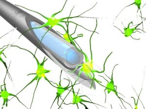
The endoscope fibres are only slightly thicker than the cells that are being studied by them.
Illustration: AG ČižmárThe research team is using ultra-thin optical fibres that are inserted into tissue to look inside the brain – but initially with test animals. Unlike conventional endoscopes, which are used for »keyhole surgery« and for diagnosing issues affecting the stomach or intestines and which consist of a bundle of fibres up to one centimetre thick, the endoscopic fibres here are only as thick as a human hair.
With a diameter of 100 micrometres, the fibres are only slightly thicker than the nerve cells themselves (see illustration on the left) and have a sharp tip that penetrates the tissue like a scalpel, causing only a minimal injury.
The key technological challenge is to guide the light through the fibres in a controlled manner. The fibres consist of a light-conducting core made of silica glass and a cladding that has a slightly lower refractive index, which causes the light to be »trapped« in the fibre and guided through it (see Light as a boundary rider). However, such »multi-mode fibres« lead to many different, unpredictable paths of light propagation, resulting in an apparently chaotic light distribution similar to that of a diffuse, opaque medium.
The researchers are sending coherent laser light through the fibre into the tissue to be examined. »Normally, the interference of light paths within the fibre core causes peaks to form in some places and troughs in others, creating a speckled ›map‹ of light and dark spots«, says physicist Čižmár. In order to obtain sharp images, the light is »pre-shaped« with a holographic modulator before it enters the fibre. This is a programmable micro-mirror array – each of the micro-mirrors can be quickly switched between two directions. »This enables us to focus the light tightly behind the fibre«, says Čižmár. Using a specific hologram and setting the micro-mirror array for each desired pixel of the image, the focus then scans the image section point by point, imaging details with a size of less than one micrometre.
There is still a long way to go before holographic endoscopy will enable insights into the human brain. However, promising results have already been obtained with test animals. Tomáš Čižmár and his research teams in Jena and Brno, Czech Republic, have already used their technology to visualize nerve cells and the cellular extensions that cells use to communicate with one another, even in deep neurological structures.
Čižmár himself started developing the methodology a good ten years ago and has focused his research on it ever since. He has received a Consolidator Grant from the European Research Council (ERC) for his »LifeGATE« project, which aims to develop light propagation and imaging technology in multi-mode fibres. »We’ve made significant progress since then«, he reports. The areas examined can now be scanned at a much higher speed, which allows greater areas to be studied overall.
In the coming years, Čižmár and his team want to use their research results to create a start-up company and offer their services to potential users in neuroscience and medicine. Their spin-off project »DeepEn« (Minimally Invasive Endoscopes for Neuroscience and Medicine) is being funded through the EXIST programme for scientific start-ups run by the Federal Ministry for Economic Affairs and Climate Action (BMWK). The spin-off is planned for the coming year. Čižmár estimates that this could lead to the development of the first diagnostic and therapeutic applications within the next ten to fifteen years.

Detail of an endoscopic image showing the processes of neurons (dendrites and axons) in the brain of a living mouse.
Image: IPHT / Institute of Scientific Instruments of the Czech Academy of Sciences