Prof. Martin Freesmeyer examines an ostrich egg in the PET/CT scanner.

The ostrich egg in the PET scanner

A nuclear medicine and chemistry research team is developing new radiodiagnostic tools for imaging in liver diseases using positron emission tomography (PET) and computed tomography (CT). The testing system is extremely unusua​l.
Prof. Martin Freesmeyer examines an ostrich egg in the PET/CT scanner.
Image: Anna Schroll/UKJ

By Uta von der Gönna

In the beginning, there was the egg—but no, this story is not quite so simple. It has at least two starting points. The egg—more precisely, the ostrich egg—only comes into the picture later.

First of all, the story is about patients with liver diseases. In order to examine a patient’s liver, we need high-resolution imaging techniques. The most commonly used technique is three-dimensional imaging in MRI (magnetic resonance imaging). Whenever an MRI scan is not possible, for example if a patient has a pacemaker or cannot tolerate contrast media because of kidney disease, the only option is scintigraphy, in which the liver and bile ducts are depicted by means of radiolabelled drugs. However, with the radiolabelled substances currently available, the optical resolution of the images leaves much to be desired. »Above all, exact three-dimensional representation, which is important for assessing organ function or for planning liver surgery, is lacking,« explains Prof. Martin Freesmeyer, head of the Clinic of Nuclear Medicine at Jena University Hospital.

The newly developed method enables detailed three-dimensional views inside the ostrich egg. Left: the egg in normal CT, right with PET.
Picture: Martin Freesmeyer/UKJ

Searching for a new contrast agent

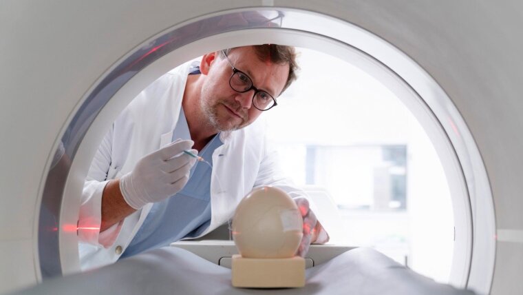
Help is found in an imaging technique that produces high-resolution three-dimensional images: positron emission tomography, or PET for short. In combination with a computed tomography scanner (CT), this method provides exact anatomical resolution and nowadays, the examination is carried out with a hybrid PET/CT scanner. Similar to the contrast agent in radiology, biomarkers coupled with radioactive isotopes (tracers) are used to detect diseases of different kinds. However, no suitable tracers have yet been established for PET/CT examinations of liver function. »The existing liver tracers are not specific enough and are extremely difficult to produce and handle,« says Prof. Freesmeyer. »Therefore, we needed a better radiodiagnostic tool for liver PET/CT.« Together with the research group of Prof. Wolfgang Weigand from the university’s Institute of Inorganic and Analytical Chemistry, the research team designed the chemical structure of several liver tracers, then synthesized and chemically characterized them.

Guiding radioactive markers precisely to the liver

The research team chose the radionuclide gallium-68 as the radioactive marker because of its relative ease of use. It can be easily obtained on-site and has a short half-life, which limits the radiation exposure of the tissue. However, the radionuclide alone does not find its way into the liver. It needs to be enveloped by specific binding molecules (»ligands«) that can guide it accurately into the liver. Fat-soluble substances are potential ligands because of their metabolism and excretion via liver cells. A template for the ligand’s structure was found in an established contrast agent for the liver, which is routinely used as a non-radioactive substance in MRI examinations.

»We equipped the ligands with additional functional groups, so that the gallium ion is sufficiently firmly bound and the substance remains stable under physiological conditions—i.e., in the blood,« says Dr Julia Greiser, describing the procedure. Greiser, a chemist, carried out these syntheses as part of her thesis and is now doing research in the Radiopharmacy Department of the Clinic for Nuclear Medicine. Like a tailor making alterations, Julia Greiser adjusted the ligands until promising candidates for the liver tracer were created.

There was also some luck involved: »The synthesis runs according to a completely new type of reaction and without elaborate reaction conditions, so that good production yields were achieved,« says Julia Greiser. The substance group and its synthesis have been patented by Prof. Freesmeyer and Dr Greiser.

New tracer undergoes testing and inspection procedures

However, before the new PET/CT tracer can be used in the clinic, it has to pass a number of tests and examinations. First, the tracer is tested to establish whether it is actually liver-specific and thus accumulates in the liver tissue after intravenous injection. Up to now, this has mostly been tested in classic animal experiments using rodents.

Apart from ethical aspects, this also has the disadvantage that expensive research equipment is required, specifically small-animal PET/CT systems. These require high investments and expensive premises, and also entail considerable expenditure on space and staff. The research team therefore looked for a solution using the existing imaging systems at the Clinic for Nuclear Medicine. Here, many patient examinations are performed daily on a PET/CT system that is significantly larger than the small-animal PET/CT. »In addition, it is important to us to carry out animal experiments only to the extent really necessary and to replace them with alternatives wherever possible,« emphasizes Prof. Freesmeyer.

Dr Thomas Winkens (r.) and medical physicist Christian Kühnel prepare an incubated ostrich egg. Part of the egg shell is removed, so that the radiolabelled tracer substance can be injected via a cannula.
Image: Anna Schroll/UKJ

Alternative to classic animal experiments

And this is where the other starting point comes into this research story. For decades, incubated hen’s eggs have been established in vaccine research, for example, and this is not regarded as a classic animal experiment. Therefore, the scientists transferred the concept of using incubated eggs to imaging and chose the largest eggs available—those of the ostrich. An ostrich egg measures about 15 centimetres in diameter and is large enough for the clinical PET/CT scanner. However, the research team was entering completely new territory when it tackled imaging using the incubated ostrich egg. »After the egg has spent 25 days in the incubator, we mark the vitelline vessels which are made visible by candling through the egg shell. Shortly before the PET/CT examination, after an incubation period of 34 to 37 days, we carefully remove a section of the calcified part of the eggshell without damaging the underlying chorion-allantois membrane with its vessels. We then inject the radiotracer using very thin cannulas, like those used in neonatology,« says nuclear medicine specialist Dr Thomas Winkens, describing the methodology.

The scientists have already obtained impressive images of the bird embryo in this way, as well as proof that the new radiotracer is highly specific to the liver. As part of a project funded by the German Research Foundation, the team will further develop the ostrich egg model in the coming years and establish it as an alternative to classic animal testing.

Preparations for patient applications

Toxicity tests are currently still being carried out in order for the new liver tracer to be used in actual patient care at some point. These tests should not be a problem, as the quantities of molecular radiotracers used are so small that they are in the »subpharmacological« range, meaning that no harmful effect is to be expected, says Prof. Freesmeyer.

The highest possible purity and precision are required in producing the tracers, for which the radioisotope is only generated shortly before the examination and then reacted with the ligands. »We are currently working on implementing the quality guidelines of drug production for the synthesis of the ligands,« explains Julia Greiser. The team is thus meeting important requirements in order to be able to use and test the new tracers on patients soon.

Information

Current Project:

In-Ovo-Imaging Using Embryonated Eggs of Large Ratite Birds—Evaluation of an Alternative to Classic Animal Research (German Research Foundation)

Contact:

Martin Freesmeyer, apl. Prof. Dr
Klinik für Nuklearmedizin
Am Klinikum 1
07747 Jena Google Maps site planExternal link